Up Block10-2 Prev Next

 上一張相片  下一張相片  索引頁
block10-2_05.jpg - Fig 1. W2254, Large intestine (cs) H&E.A cross section through the colon is shown at low magnification.It shows the four layers that make up the wall of the colon: themucosa (M), the submucosa (S), the muscularis externa (ME),and the serosa (Se). Although these layers are the same asthose in the small intestine, several differences should be noted.The large intestine has neither villi nor the plicae circulares. Onthe other hand, the longitudinal layer of the muscularis externais substantially thinner than the circular layer except in threelocations where the longitudinal layer of smooth muscle ispresent as a thick band, called teniae coli (TC).

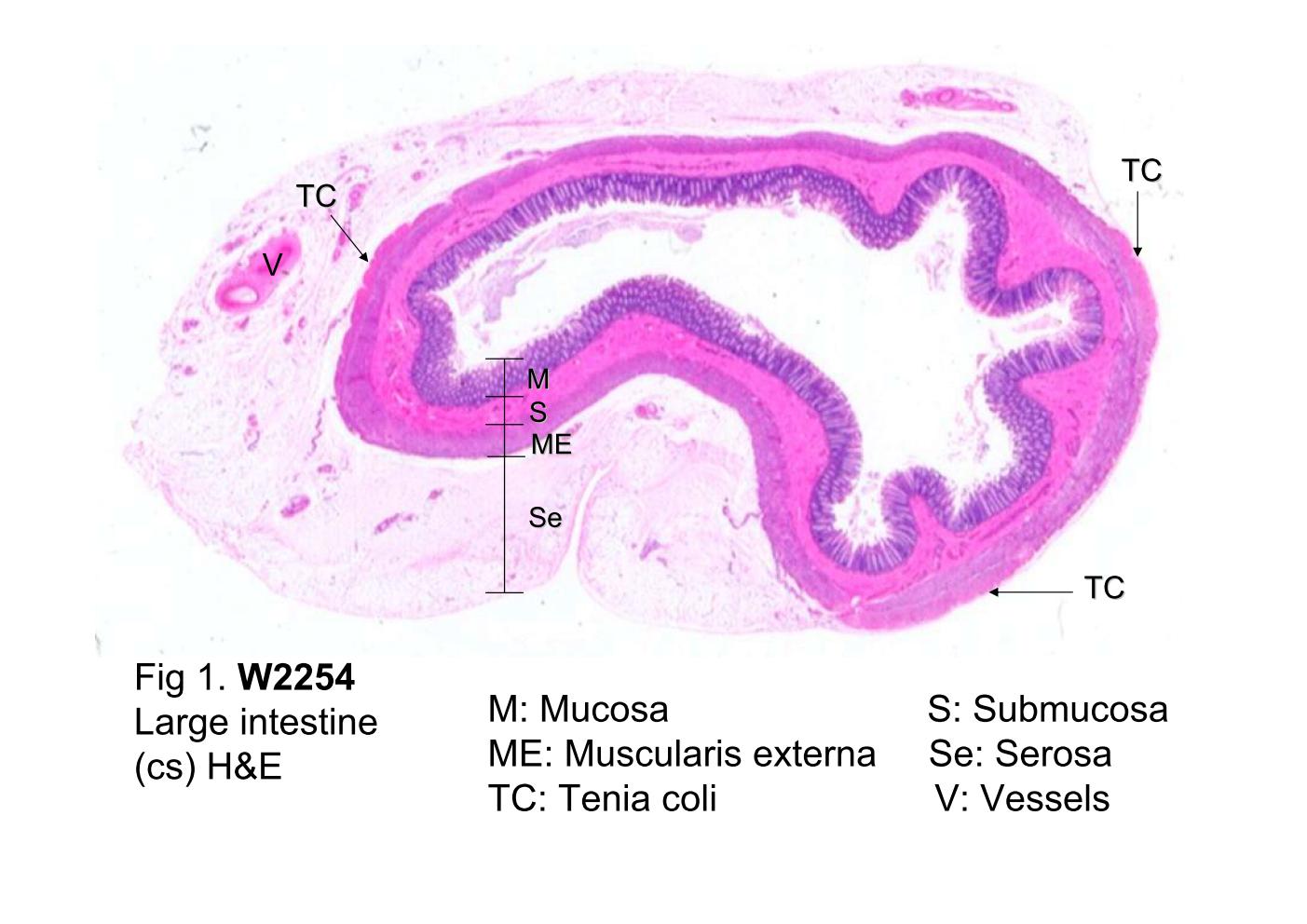
Fig 1. W2254, Large intestine (cs) H&E. A cross section through the colon is shown at low magnification. It shows the four layers that make up the wall of the colon: the mucosa (M), the submucosa (S), the muscularis externa (ME), and the serosa (Se). Although these layers are the same as those in the small intestine, several differences should be noted. The large intestine has neither villi nor the plicae circulares. On the other hand, the longitudinal layer of the muscularis externa is substantially thinner than the circular layer except in three locations where the longitudinal layer of smooth muscle is present as a thick band, called teniae coli (TC).
 block10-2_01block10-2_02block10-2_03block10-2_04block10-2_05block10-2_07block10-2_09block10-2_11block10-2_13block10-2_15

支援訊息