Up Block7 Prev Next

 上一張相片  下一張相片  索引頁
block7_05.jpg - Fig. 1. Micrograph of multipolar neurons. Multipolar neuronswere smear with phloxine staining and counterstain with themethylene blue for neuronal nucleus. A typical multipolarneuron have multiple processes and surrounded by thenumerous supporting cells.

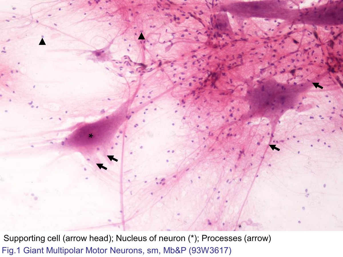
Fig. 1. Micrograph of multipolar neurons. Multipolar neurons were smear with phloxine staining and counterstain with the methylene blue for neuronal nucleus. A typical multipolar neuron have multiple processes and surrounded by the numerous supporting cells.
 block7_01block7_02block7_03block7_04block7_05block7_07block7_09block7_11block7_13block7_14

支援訊息